Intravascular Ultrasound (IVUS) is an advanced medical imaging technique that allows cardiologists to look inside blood vessels, especially the coronary arteries. Unlike traditional imaging, IVUS uses high-frequency sound waves to create a detailed cross-sectional image of the vessel walls.
Doctors mainly use IVUS during angioplasty or stent placement to assess plaque build-up, measure artery size, and guide precise treatment.
History of IVUS
- First introduced in the 1980s as a research tool.
- By the 1990s, IVUS started being used in cardiac catheterization labs worldwide.
- Over time, advancements in catheter design and high-resolution imaging made IVUS more reliable and accessible.
- Today, IVUS is considered a gold standard imaging method alongside Optical Coherence Tomography (OCT).
Table of Contents
How IVUS Works?
IVUS works on the principle of ultrasound imaging but inside blood vessels.
Step-by-Step Process:
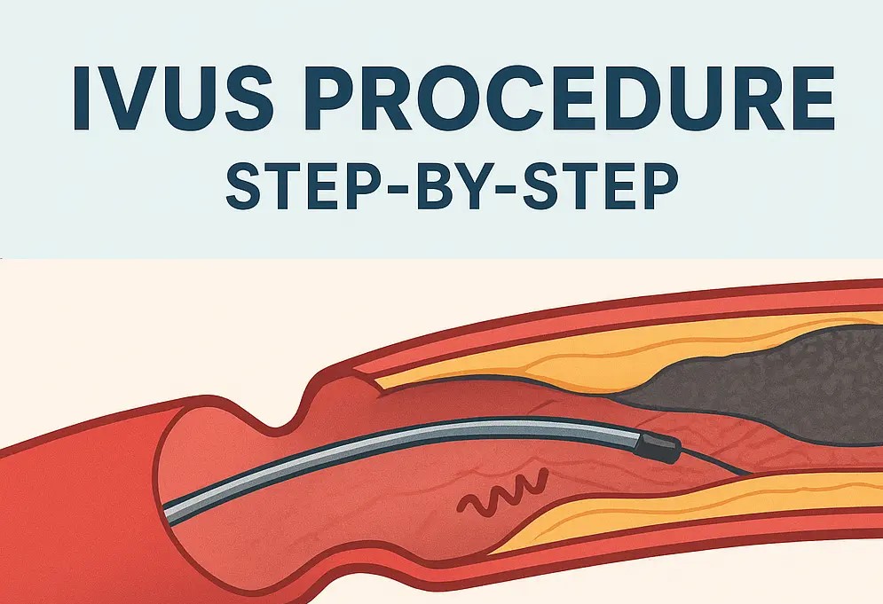
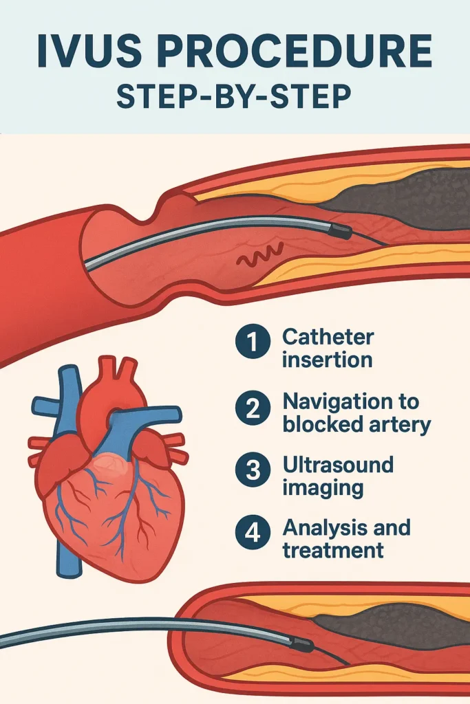
- Catheter Insertion: A thin IVUS catheter is inserted through an artery, usually from the groin or wrist.
- Navigation: The catheter is guided to the blocked or narrowed section of the artery.
- Ultrasound Emission: The transducer at the catheter tip emits high-frequency sound waves.
- Echo Reception: The echoes return from vessel walls and are captured by the device.
- Image Formation: A real-time cross-sectional image of the artery is displayed on the IVUS monitor.
- Diagnosis & Treatment: The cardiologist measures artery size, plaque thickness, and guides stent placement.
Types of IVUS Systems
There are mainly two types of IVUS imaging systems:
- Mechanical IVUS
- Uses a single rotating transducer.
- Produces high-resolution images.
- Slightly slower.
- Phased-Array IVUS
- Uses multiple transducers arranged in an array.
- Faster and allows automated pullback imaging.
- More common in modern cath labs.
Some advanced IVUS machines also integrate Virtual Histology (VH-IVUS) to analyze plaque composition.
IVUS Procedure in Cath Lab – Step by Step
The IVUS procedure is usually performed during angioplasty or stent placement.
- Preparation: Patient is given mild anesthesia and connected to monitoring systems.
- Catheter Insertion: A guidewire is placed in the artery, followed by the IVUS catheter.
- Imaging: The catheter moves through the artery, capturing images in real-time.
- Analysis: Cardiologists check plaque size, vessel diameter, and blockage severity.
- Treatment: Based on IVUS results, stents are placed with better accuracy.
- Completion: The catheter is removed, and the site is bandaged.
The whole process usually takes 30–45 minutes.
IVUS vs Angiography
| Feature | IVUS | Angiography |
|---|---|---|
| Imaging | Cross-sectional, detailed | 2D shadow images |
| Accuracy | High (shows plaque composition) | Limited |
| Radiation | No radiation (ultrasound) | Uses X-ray |
| Cost | Higher | Lower |
| Use Case | Stent guidance, plaque analysis | Basic artery block detection |
👉 IVUS is more detailed, while angiography is faster and cheaper. Many cardiologists use them together.
IVUS Catheter Explained
The IVUS catheter is the most critical component.
- It is a thin, flexible tube with an ultrasound transducer at its tip.
- Diameter: Usually 2.9F to 3.5F.
- Function: Emits sound waves and captures echoes.
- Designed for smooth navigation inside small coronary arteries.
IVUS Machine and Components
An IVUS system includes:
- Console: Controls imaging, records data.
- Software: Processes ultrasound echoes into images.
- Catheter: For inside-vessel imaging.
- Pullback Device: Moves catheter at constant speed for detailed scans.
Clinical Uses of IVUS
- Diagnosing arterial blockages
- Guiding stent placement
- Measuring vessel diameter
- Plaque composition analysis
- Checking stent expansion after placement
- Reducing risk of restenosis (re-blockage)
IVUS in Cardiology
IVUS is widely used in coronary interventions, especially when angiography results are unclear. It helps cardiologists decide:
- Whether a stent is required
- What size of stent to use
- How well the stent is placed
This makes angioplasty safer and more effective.
Cost of IVUS Procedure
The cost of IVUS depends on country, hospital, and case complexity.
- India: ₹40,000 – ₹70,000 per procedure
- USA: $2,000 – $4,000
- Europe: €1,500 – €3,000
Advantages of IVUS
- High-resolution imaging
- Helps select correct stent size
- Detects hidden plaque not seen on angiography
- No radiation risk
- Reduces complications during angioplasty
Risks and Limitations
- Slight risk of artery damage
- Higher cost compared to angiography
- Requires specialized training
- Limited availability in smaller hospitals
Post-IVUS Care
- Patients may feel mild discomfort at the catheter site.
- Avoid heavy lifting for 1–2 days.
- Continue prescribed medicines.
- Follow-up checkups with cardiologist recommended.
Future of IVUS
- Integration with Artificial Intelligence (AI) for automated diagnosis.
- 3D IVUS imaging for more accurate visualization.
- Combined IVUS + OCT imaging systems.
FAQs about IVUS
1. What is IVUS in cardiology?
IVUS is a catheter-based ultrasound system used to view inside coronary arteries.
2. How long does an IVUS procedure take?
Usually 30–45 minutes, depending on the case.
3. Is IVUS better than angiography?
Yes, IVUS provides detailed cross-sectional images, while angiography only shows shadows.
4. What is the cost of IVUS in India?
Around ₹40,000 – ₹70,000 depending on hospital.
5. Is IVUS safe?
Yes, it is generally safe, with minimal risks.
6. Can IVUS replace angiography?
No, but it complements angiography for better accuracy.